+
19
M
Confirmed cases
Loading...
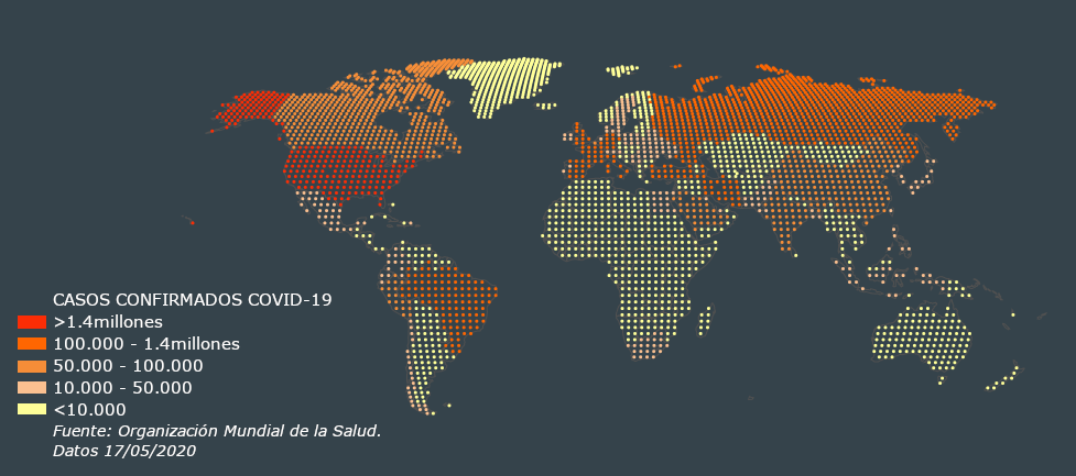
COVID-19 is an infectious disease caused by the SARS-CoV-2 virus, which can be transmitted before the appearance of symptoms (presymptomatic) or even if the person has no symptoms (asymptomatic). This together with a contagion rate, estimated by the WHO, of 1.4 to 2.5, is what has led to the COVID-19 being classified as a pandemic.
The virus mainly affects the airways starting at the throat and continuing through the bronchial tubes to the lungs. To fight the virus, the body has an inflammatory response that can damage the lungs. This is why medical images have played an important role in the absence of tests to detect the disease.
Confirmed cases
Healed patients
Deaths from SARS-CoV-2
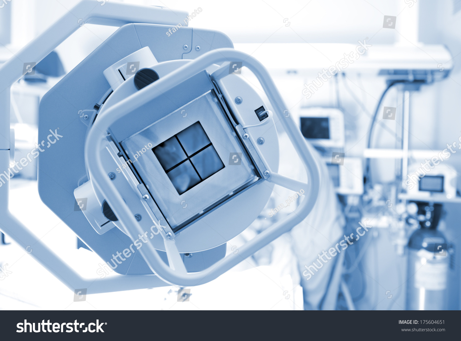
Chest X-Ray is a common, non-invasive and rapid test, widely used by clinicians worldwide to diagnose all sorts of respiratory diseases and viral infections affecting the respiratory system. Given the lack of PCR tests and the saturation of health systems, X-rays have been used as a triage tool 1 for patients who come to health systems for health care and as a diagnostic tool to monitor the accelerated progression of lung abnormalities in infected patients
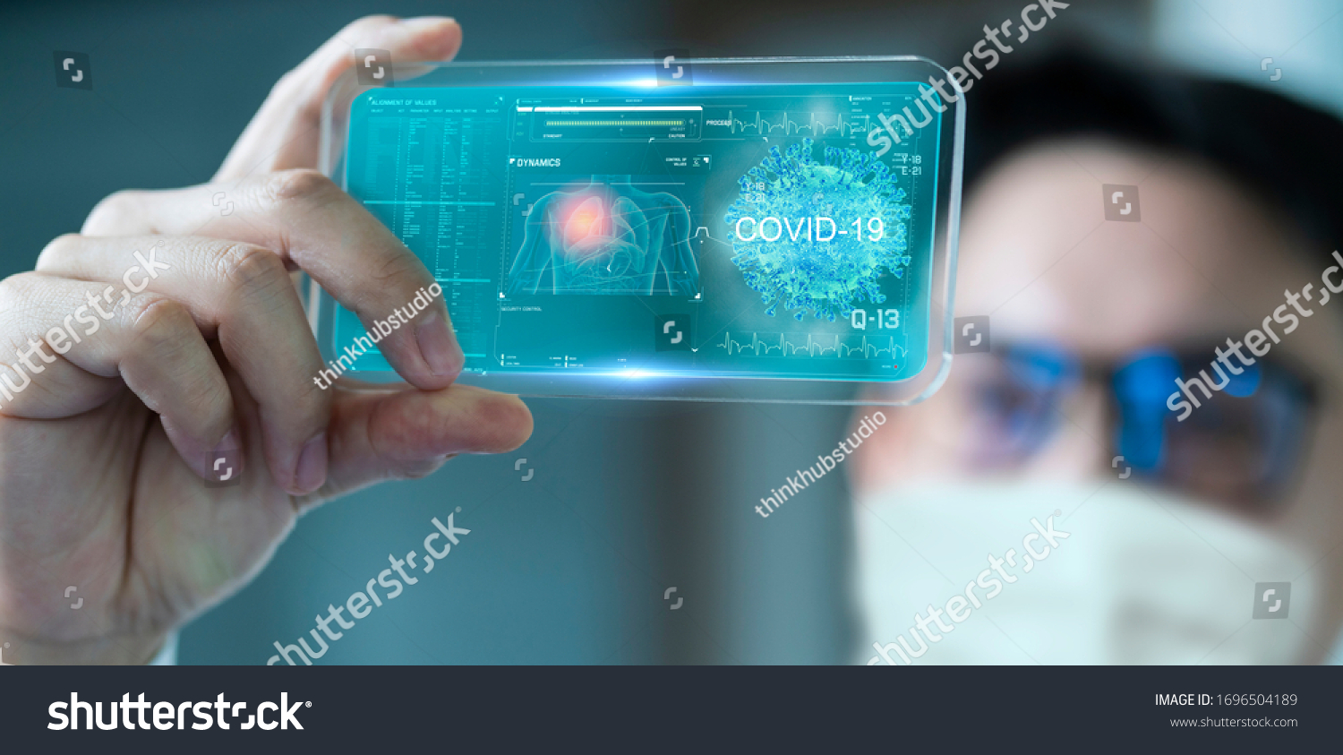
Due to the exponential increase in patients, medical personnel have been overwhelmed, which is why we need to find ways to help them fight this disease. These days, medical imaging has been key to triage of patients in the emergency department, but it must be borne in mind that the evaluation of an image by a radiologist takes about 15 minutes. This is where the AI intervenes reducing these times since the processing of an image takes a few seconds.
Therefore AI allows reducing the workload on doctors in a scenario like the current outbreak of COVID-19.
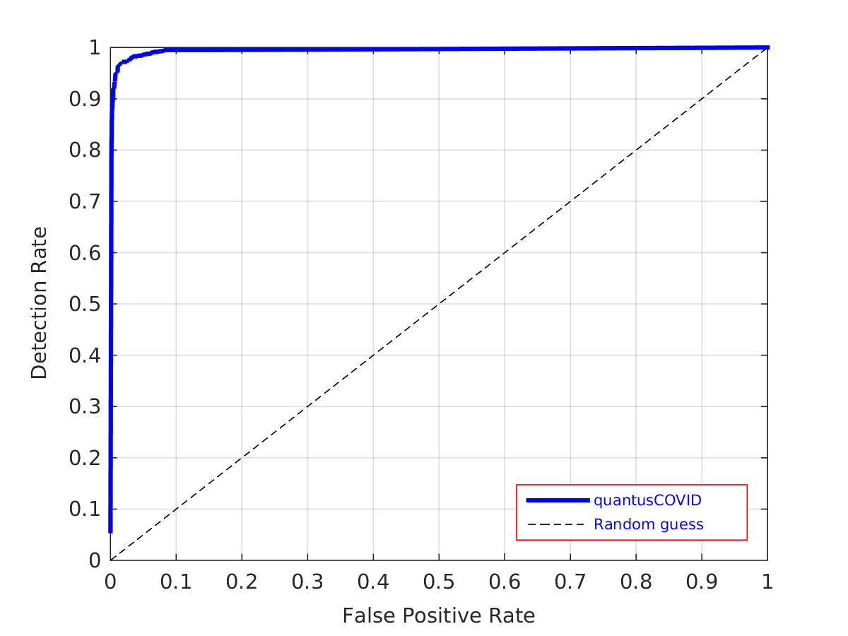
In a first study, tested on 3,528 patients,the results indicate that the texture analysis using Deep Learning techniques provides information
associated with COVID-19 involvement in CT images. With 96.4% global success and a 1% false positive rate.
The study, "Computer-aided covid-19 patient
screening using chest images (X-Ray and CT scans)" 3 was published in MedrXiv.
The next step is to have a larger database to corroborate that the results obtained are still valid and in this way be able to offer
quantusCOVID19 to the world.


quantusCOVID19 is a service that is available 24 hours a day, 365 days a year.
El pago de la Prueba de Madurez Pulmonar Fetal ha sido cancelado.
Cierre esta ventana e inténtelo de nuevo.
Puede contactar con nosotros en el teléfono +34 626667989 o en el email support@quantusflm.com
El pago de su test quantusFLM ha sido realizado correctamente.
En breve recibirá un correo electrónico con el código de la prueba.
Debe marcar que acepta los Términos y Condiciones de compra antes de efectuar el pago.
| Nombre: (*) | |
| Apellidos: (*) | |
| Teléfono: | |
| eMail: (*) | |
| Id. de quantusFLM: (*) | |
|
¿No te acuerdas de tu usuario?. |
| eMail: (*) |
The terms and conditions contained in this Legal Notice regulate the use of the website www.quantuscovid19.org (hereinafter, the "Website") that TRANSMURAL BIOTECH SL (hereinafter, "TRANSMURAL BIOTECH") makes available to users Accessing your Website (hereinafter, the "User" or "Users") in order to provide them with information about their medical testing services, as well as the possibility of using them (hereinafter, the "Services").
TRANSMURAL BIOTECH SL, with CIF number B-65084675, has its registered office at Beethoven 15 Floor 4 Office 18 08021-Barcelona, Spain, and is registered in the Mercantile Register of Barcelona, in Volume 41.169, Folio 180, Page B-381404 , 1st Registration. If you wish to contact TRANSMURAL BIOTECH you can use the postal address indicated above, through the telephone number or if you prefer, through the email address info@transmuralbiotech.com
The use of the Website by Users implies the acceptance of all the conditions included in this Legal Notice and the Privacy Policy.
The provision of the Website Services will have a limited duration at the moment in which the Users are connected to the Website or any of the Services that are provided through it. Therefore, Users must carefully read the Legal Notice present on the Website each time they intend to use it, as it may undergo modifications without prior notice.
TRANSMURAL BIOTECH informs you that the Assistance Services offered through the Website are subject to the Terms of Contract that the User, before the purchase, must accept. The Terms of Contract, replace, complete and / or modify, if applicable, these Conditions of Use.
Access to and / or use of said Services and Content (as defined in Clause 7 of this Legal Notice), expresses the entire and unconditional acceptance of the particular conditions in the version published by TRANSMURAL BIOTECH at the time it occurs. said access and / or use.
It is possible that, throughout the provision of the Services included in the Website, the name of the domain under which said Services are provided will be modified. Therefore, the Users know and accept this possibility, remaining in such case, fully valid the obligations accepted by the Users and TRANSMURAL BIOTECH according to what is established in this document.
Your use of the Website and its Contents and Services is entirely voluntary and remains under your sole responsibility.
Access to the Website is free except for the cost of the connection through the telecommunications network provided by the access provider contracted by the User. Certain Services are exclusive to certain Users and their access is restricted.
The Users can not under any circumstances modify or delete the identifying data that exist in their case of the rights of TRANSMURAL BIOTECH or of third parties. The Users may only access the Content and Services through the means or procedures that have been made available to this effect on the Website or are routinely used on the Internet for that purpose, provided that they do not imply the violation of intellectual property rights or industrial or imply some type of damage to the Website and / or its information or Contents and / or Services offered.
The Users are obliged to use the Contents and Services in a diligent, legal, correct and lawful manner and, in particular, they commit themselves to a merely enunciative and non exhaustive title to abstain from:
1. use the Contents and Services in a manner and for purposes contrary to law, morality and generally accepted good practices or public order;
2. use the Contents and Services in such a way that they produce or may produce effects contrary to the law, morals and generally accepted good practices or public order;
3. transmit or disseminate information, data, content, messages, graphics, drawings, sound and / or image files, photographs, recordings, software, and in general, any material that is obscene, offensive, vulgar or that induces criminal, slanderous, defamatory, infamous, violent or, in general, contrary to the law, morals and generally accepted good customs or public order;
4. reproduce, copy, or distribute the Contents, as well as allowing public access to them through any form of public communication, or transform or modify them, unless the authorization of the owner of the corresponding rights or it is legally permitted;
5. Violate intellectual or industrial property rights belonging to TRANSMURAL BIOTECH or third parties;
6. use the Services and Content in a way that may cause damage or overload to the operation of the Website;
7. perform fraudulent transactions or that may facilitate illegal or fraudulent behavior of any kind;
8. Use the Content and Services and, in particular, the information of any kind obtained through the Website with any type of advertising purpose and, in particular, to send advertising, communications for the purpose of direct sale or with any other kind of advertising, commercial purpose, unsolicited messages individualized or directed to a plurality of people, as well as to commercialize or disclose in any way said information.
Users will be liable for damages of any nature that TRANSMURAL BIOTECH may suffer, directly or indirectly, as a consequence of the breach of any of the obligations derived from this Legal Notice or the law in relation to the use of the Website and the Contents and / or Services.
The Content available on the Website is provided with the understanding that TRANSMURAL BIOTECH is not dedicated to providing medical, therapeutic or any other type of professional advice or service. These Contents are provided exclusively as general help for instructional purposes and should not be considered as medical or health advice or used to diagnose or treat specific problems. The Contents are not intended to replace the advice or professional services of a doctor who is familiar with the specific circumstances of the User. Always consult your doctor or medical company regarding any illness and before undergoing any new treatment.
TRANSMURAL BIOTECH reserves the right to interrupt access to the Website, as well as the provision of any or all of the Content and / or Services provided through it at any time and without prior notice, whether for technical reasons, security, control, maintenance, power failure or any other cause.
Consequently, TRANSMURAL BIOTECH does not guarantee the reliability, availability or continuity of the Website or the Contents and / or Services, so the use thereof by Users is carried out at your own risk, without that, at any time, responsibilities can be demanded for the discontinuity or lack of availability of the Contents and / or its Services.
TRANSMURAL BIOTECH will not be liable in case of service interruptions, delays, errors, malfunctions and, in general, other inconveniences that have their origin in causes beyond the control of TRANSMURAL BIOTECH, and / or due to a performance fraudulent or negligent of the Users and / or have causes of Force Majeure. Without prejudice to what is established in article 1105 of the Civil Code, the concept of Force Majeure will also be understood, and for the purposes of these general conditions, all events that occurred outside the control of TRANSMURAL BIOTECH, such as: third parties, operators or service companies, lack of access to third party networks, acts or omissions of the Public Authorities, those others produced as a result of natural phenomena, blackouts, etc. and the attack of hackers, crackers or other third parties on the security or integrity of the computer system. In any case, whatever its cause, TRANSMURAL BIOTECH will not assume any responsibility for direct or indirect damages, consequential damages and / or loss of profits.
TRANSMURAL BIOTECH excludes any liability for damages of any kind that may be due to the lack of veracity, accuracy, completeness and / or timeliness of the Contents transmitted, disseminated, stored, made available or received, obtained or to which accessed through the Website, nor by the content provided or offered by third parties or entities. TRANSMURAL BIOTECH will try as much as possible to update and rectify that information hosted on the Website that does not comply with the minimum guarantees of truthfulness. However, it will be exonerated from responsibility for its non-updating or rectification as well as for the Contents and information provided in it.
TRANSMURAL BIOTECH is not responsible for the Content of the information collected on the Website, as well as for those opinions, comments, judgments or any other manifestation contained in it that are not issued directly by TRANSMURAL BIOTECH.
TRANSMURAL BIOTECH excludes any liability for damages of any kind that may be due to the presence of viruses or the presence of other harmful elements in the Contents that may cause alteration in the computer systems, as well as in the documents or systems stored in the same.
TRANSMURAL BIOTECH is not responsible for the Contents, whatever they may be, that Users send to TRANSMURAL BIOTECH through the Website, through the electronic mail service or by any other means, thus being attributable to Users any responsibility arising from the Content sent by them.
TRANSMURAL BIOTECH is not responsible for the use that Users make of the Website Content, as well as any other material contained on the Website, which may involve a violation of any type of national or international standard of rights of intellectual or industrial property or any other right of third parties. Similarly, it is not responsible for possible security errors that may occur due to the use of non-updated versions of browsers, or the consequences that may arise from the malfunctioning of the browser, due to improper configuration, presence of computer viruses or any other cause beyond TRANSMURAL BIOTECH.
TRANSMURAL BIOTECH guarantees that it has signed agreements with the Centers or Medical Professionals that make up the Medical Board to provide the medical service to the User. TRANSMURAL BIOTECH will not be responsible for delays, defective fulfillments or breaches in the provision of the Medical Service when they were due to the failure of the Center or Medical Professional. In particular, in relation to the professional medical activity itself, the diagnoses and treatments to the Users, TRANSMURAL BIOTECH will be totally alien and will not have any responsibility. The Center or Medical Professional will, therefore, be solely responsible for any obligation, right or result derived from the provision of its services.
The access service to the Website may include technical link devices, directories and even search tools that allow Users to access Internet websites (hereinafter, "Linked Sites"). In these cases, TRANSMURAL BIOTECH acts as a service provider in accordance with article 17 of Law 34/2002, of July 11, on Services of the Information Society and Electronic Commerce (hereinafter, the "LSSI") and will only be responsible for the contents and services provided in the Linked Sites to the extent that it has actual knowledge of the illegality or that may be injured goods or rights of third parties and has not deactivated the link with due diligence.
In the event that Users consider that there is a Linked Site with illicit or inappropriate content, they may communicate it to TRANSMURAL BIOTECH at the contact address indicated in the first section of this Legal Notice, indicating:
1. the caller: name, address, telephone number and email address;
2. a description of the facts that reveal the illicit or inappropriate nature of the Linked Site;
3. an express statement that the information contained in the communication is accurate. In no case does this communication entail the obligation to withdraw the corresponding link, nor does it imply, according to the provisions of the LSSI, the effective knowledge of the activities and / or contents indicated by the communicator.
In any case, the existence of Linked Sites must presuppose the existence of agreements with the managers or owners thereof, or the recommendation, promotion or identification of TRANSMURAL BIOTECH with the statements, content or services provided.
TRANSMURAL BIOTECH does not know the contents and services of the Linked Sites and therefore is not responsible for the damages caused by the illegality, quality, outdated, unavailability, error and uselessness of the contents and / or services of the Linked Sites or by any other damage that is not directly attributable to TRANSMURAL BIOTECH, except as provided in the aforementioned article 17 of the LSSI.
If Users decide to visit and / or use any Linked Sites, they will do so at their own risk, and they will have to take appropriate protection measures against viruses or other harmful elements.
The Internet user who wants to introduce links from their own web pages to the Website must comply with the conditions set out below without ignoring the responsibilities derived from the law:
1. The link will only link to the home page or main page of the Website but will not be able to reproduce it in any way (inline links, deep-links, browser or border enviroment, copy of texts, graphics, etc.);
2. It will be prohibited in any case, in accordance with the applicable legislation and in force at any time, to establish frames or frames of any kind that involve the Website or allow the display of the Contents through Internet addresses other than those of the Site. Web and, in any case, when viewed together with contents external to the Website in such a way that: (i) it produces, or may produce, error, confusion or deception in the Users about the true origin of the Service or Content; (ii) suppose an act of comparison or unfair imitation; (iii) serves to take advantage of the reputation of the brand and prestige of TRANSMURAL BIOTECH; or (iv) in any other way is prohibited by current legislation;
3. Any type of false, inaccurate or incorrect manifestation or indication about TRANSMURAL BIOTECH, employees, customers or about the quality of the Services provided will not be made from the website that introduces the link;
4. In no case, will be expressed or implied on the web page where the link is located that TRANSMURAL BIOTECH has given its consent for the insertion of the link or that otherwise sponsors, collaborates, verifies or supervises the services of the sender;
5. The use of any word, graphic or mixed mark or any other distinctive sign of TRANSMURAL BIOTECH within the sender's website is prohibited except in cases permitted by law or expressly authorized by TRANSMURAL BIOTECH and whenever allowed, in these cases, a direct link to the Website in the manner established in this clause;
6. The web page that establishes the link must faithfully comply with the law and can not under any circumstances dispose of or link to its own or third party content that:
1. are illegal, harmful or contrary to morals and morals (pornographic, violent, racist, etc.);
2. induce or may induce in the User the false conception that TRANSMURAL BIOTECH subscribes, endorses, adheres to or in any way supports the ideas, statements or expressions, legal or illegal, of the sender;
3. they are inappropriate or not pertinent with the activity of TRANSMURAL BIOTECH in attention to the place, contents and theme of the web page of the sender;
The establishment of the link does not imply in any case the existence of relations between TRANSMURAL BIOTECH, and the owner of the website on which it is established, nor the acceptance and approval by TRANSMURAL BIOTECH of its contents or services offered therein made available of the public.
TRANSMURAL BIOTECH may request, at any time and without the need to provide the reasons for such request, that any link to the Website be removed, after which the person responsible for the linking website must proceed immediately to its elimination.
The entire content of the Website, understood by these as merely enunciative texts, photographs, graphics, images, icons, technology, software, links, domains, brands and other audiovisual or sound content, as well as its graphic design and codes source, are the exclusive property of TRANSMURAL BIOTECH or third parties, whose rights, if any, are recognized by TRANSMURAL BIOTECH, and are subject to intellectual and industrial property rights protected by national and international legislation (hereinafter, the "Contents").
It is strictly forbidden any use of any of the elements subject to industrial and intellectual property with any type of purpose, especially commercial, as well as its distribution, public communication, modification, alteration, transformation or decompilation, unless expressly authorized in writing by the owner of them.
The infringement of any of the aforementioned rights may constitute a violation of these provisions, as well as an action constituting an offense established in articles 270 and following of the Penal Code.
Last modified: January 16th , 2019
TRANSMURAL BIOTECH SL. (hereinafter, "TRANSMURAL"), with CIF number B-65084675, with registered office at Beethoven 15 Floor 4 Office 18, 08021-Barcelona, and registered in the Mercantile Registry of Barcelona, in Volume 41.169, Folio 180, Sheet B- 381404, Registration 1 is the owner of the website https://www.quantusflm.com (hereinafter, the "Website").
The Terms of Contract are available to all Users of the Website from the link Terms of Contract located in the footer of the "Website" freely and free.
If the User wishes to contact TRANSMURAL for any doubt or incident with his purchase, he can contact through the telephone number +34 931190229 from 09:00 to 18:00 (Spain time), or if you prefer by email at the address info@transmuralbiotech.com
The contract between TRANSMURAL and the Client is understood to be perfected from the moment in which it concludes the contracting procedure by pressing the "Buy" button, it being understood that the monitoring of all phases of the electronic contracting procedure and the inclusion of all requested data suppose, together with the marking of the corresponding box relative to the acceptance of these Terms of Contract, a direct manifestation of the Client's willingness to accept them.
The language in which the contracting procedure will be processed and in which this contract is formalized will be, unless otherwise indicated, Spanish.
TRANSMURAL reserves the right to unilaterally modify these Terms of Contract at any time. All modifications of the Terms of Contract will be published on the Website. The Client will be subject to that version of the present document that he accepted at the time of purchase.
The date from which these Terms of Contract are in force is the date at the beginning of the document
The procedure for contracting the offered products is carried out electronically through the Website. The complete procedure to be followed by all Users wishing to acquire any of the products and / or services offered through the Website will be as follows:
Once the User has accessed the Website, they must select those products and / or services that interest them, review their descriptions, as well as their characteristics, conditions, what is included and not included, and final prices indicated in the descriptive sheet of each product and / or service.
Then the User must read and expressly accept these Terms of Contract, as well as the Privacy Policy, by marking the corresponding boxes provided for that purpose and clicking on the "Buy Now" button.
Payment is made through the REDSYS System, the user will be redirected, automatically by the system to the online payment platform (TPV) of the corresponding banking entities.
The User must enter the payment information requested and complete the electronic purchase process, for which you must only click on the "Pay Now" button.
After the purchase, a summary screen of the purchase will be displayed, notwithstanding that the Customer automatically receives an email confirming that the purchase has been made successfully.
This e-mail will describe the purchase made, as well as the characteristics thereof. This document will serve as accreditation for any type of claim. In case of not receiving such email, the Client should check his "spam" or "spam" tray and, if he is not in that section, he should notify TRANSMURAL in the shortest time possible so that the incident.
All prices shown on the Website are final prices of the products and / or services. These prices will be shown in Euros (€). In the event that the final price changes due to increases or discounts that are applicable, expenses passed on to the Customer and / or additional expenses for products and / or accessory services, means of payment, etc., all these amounts will be shown to the Client. They will be shown broken down and prior to payment during the contracting process.
The payment of the Assistance Service is made through a credit or debit card and will be charged to the Client's account at the time of purchase. To proceed with the payment, the Customer must follow each and every one of the instructions indicated at the time of purchase, providing the following information: Name of the cardholder, type of card, card number, expiration date of the card or any other that is required during the purchase process. The Client undertakes not to provide false information, including names, addresses and / or contact or payment details, as well as not to initiate any illegal activity in connection with the purchase and not to allow anyone to do so.
To make the payment, the Client will be redirected to the to the online payment platform of the corresponding banking entities, where he can pay with his debit or credit card in a secure way.
TRANSMURAL states that it does not have access to or store sensitive data relative to the means of payment used by the Client. Only the corresponding entity processing the payment has access to these data as a way of managing payments and collections.
All data provided for these purposes are encrypted to ensure maximum security of them.
In accordance with the provisions of current regulations regarding consumers and users, the Client may, without needing to justify his decision and without any penalty, exercise the right of withdrawal during the fourteen (14) calendar days following the date of purchase, as long as you have not used the Service.
In order to exercise the right of withdrawal, the Client must state this by sending an unambiguous communication to the email address of TRANSMURAL indicated in these Terms of Contract indicating:
- Name and surname of the Client;
- Authorization code;
- Email address used for the purchase.
TRANSMURAL will communicate by e-mail the acknowledgment of receipt of the Customer's withdrawal and will make the refund of the amount of the Code within a maximum period of fourteen (14) calendar days from the day of request of withdrawal.
Any refund will be made through the same means of payment with which the Client made the transaction. In the event that the means of payment has been canceled, expired or has ceased to be valid for any reason, the Customer must notify it to the e-mail address infor@transmuralbiotech.com. Otherwise, TRANSMURAL will not be responsible for the refund, so the Client must contact his bank or payment service provider to process the refund.
The refund will be considered executed if the Client does not reject it within a period of fourteen (14) calendar days from the date of receipt. The exercise of the right of withdrawal will extinguish the obligations of TRANSMURAL with the Client in relation to the Code and / or the Assistance Service on which he has withdrawn.
TRANSMURAL guarantees that the contents, data or information related to the products and / or services offered on its Website are reliable, truthful and accurate. However, TRANSMURAL will not be responsible for those contents, data or information that have been introduced, displayed or modified by third parties outside TRANSMURAL.
The photographs, texts, graphics, information or other content and characteristics reproduced that illustrate the products and / or services marketed by TRANSMURAL are merely illustrative, so they may vary. Notwithstanding the foregoing, TRANSMURAL will use its best efforts to make the description of the products and / or services as close as possible to reality.
Both the Client and TRANSMURAL undertake to comply with their legal and contractual obligations generated by virtue of this contract.
TRANSMURAL will not be responsible in case of unavailability of the products and / or services offered through the Website when it is due to force majeure, theft or loss, or error in the order or data provided by the User.
TRANSMURAL will use all efforts at its disposal to keep products and / or services available through the Website, except for lack of availability or performance due to:
Inactivity or temporary unavailability of the Website due to updating and / or technical maintenance thereof, of what will be previously reported, and as soon as possible, through publication on the Website itself.
Causes beyond the control of TRANSMURAL, such as force majeure, problems of access to the Internet, technological problems, actions or omissions of third parties, etc.
In all the aforementioned cases, beyond the control and due diligence by TRANSMURAL, the User will not be entitled to any compensation from TRANSMURAL for damages, direct or indirect, or for loss of profit.
In case of closure or suspension of the Website for reasons beyond the control of TRANSMURAL, and whenever possible, the User will be promptly informed of the transfer of the service to a new domain, modifying only the stipulations of these Terms of Contract as relative to the domain in which the Website remains active.
If the Client uses the Code, but the Healthcare Provider does not provide the Healthcare Service adequately or if he / she has any complaint or comment regarding the execution of the Healthcare Service, he / she must submit his claim directly to the Healthcare Provider, and TRANSMURAL is not entitled to receive it of no claim.
Except as otherwise provided in the applicable legislation, for any litigious matter or matter pertaining to the Website or the contracting of products and / or services regulated in these Terms of Contract, Spanish law shall apply.
Likewise, in the case of disputes related to online contracting, we inform you that the European Commission provides consumers with an Online Claims Resolution Platform for consumer matters, which can be accessed through the following link
These terms and conditions (hereinafter, the "Terms of Contract") regulate the intermediation service through which TRANSMURAL makes available to the Client, for contracting, the products and / or services delivered and / or provided by the Care Provider to the Customer (hereinafter, the "Assistance Services") once the latter submits the Authorization Code.
The Terms of Contract are available to all Users of the Website from the link Terms of Contract located in the footer of the "Website" freely and free.
By "Code" shall be understood, for the purposes of these Terms of Contract, the electronic or physical instrument that empowers the owner thereof (hereinafter, the "Client") to receive the Assistance Service.
The medical or welfare professional is a third party that is not part of TRANSMURAL and delivers and / or provides the Assistance Service hired by the Client by delivering the Code (hereinafter, the "Care Provider").
The Client must verify before the purchase that there is one in the list of Assistance Providers where he can go to take the test.
The Customer, when purchasing the Care Service, will receive by email the Code that must be delivered to the Care Provider. Then, the Customer must call the medical center of the chosen Care Provider to book an appointment. The Code gives the right to the provision of the Healthcare Service acquired.
Unless otherwise indicated, the Code:
1. can only be redeemed once;
2. can only be used for the contracted service.
Therefore, the purchase of a Code is understood to be made for personal use, either by the Client or by any third party designated by it. It is expressly prohibited:
1. The sale or use for lucrative purposes of the Code.
2. The copy of the Code.
Once the Code has been issued, the Client will be solely responsible for its custody. Neither TRANSMURAL nor the Care Provider is responsible for the loss or theft of the Code. The Authorization Code is under the sole responsibility of the Client.
The use of the Code for purposes other than those provided in these Terms of Contract (i) may lead to the cancellation of the Code, and (ii) in any case exempt TRANSMURAL from any liability to the Client and to the third party purchaser or any third party, also reserving TRANSMURAL how many actions correspond to it in law.
If the Client wishes to hire additional tests, he must acquire these benefits before attending the consultation. If you do not enforce the Code within the Validity Period, the Code will automatically expire and will be rendered ineffective, so that the Customer will not be entitled to any refund.
Unless the Healthcare Service itself allows it, the Code does not entitle the holder to receive the Care Service at a certain time, so the Client must agree with the Care Provider when providing the Care Service.
The Code can only be exchanged completely. No exchanges or partial cancellations will be accepted. If you agree with the Care Provider the exchange of the Code for a service of lesser value of the contracted Healthcare Service, the Client will not be entitled to the refund of the remaining money, nor to have credit or a new Code equivalent in value to the difference between the original value and the value of the Assistance Service enjoyed.
If the Care Provider selected by the Client cannot provide the Healthcare Service under the agreed conditions, TRANSMURAL will make available to the Customer the refund of the purchase price of the Code.
TRANSMURAL will not be responsible in case of unavailability of the products and / or services offered through the Website when it is due to force majeure, theft or loss, or error in the order or data provided by the User, as well as in case of errors, actions or omissions attributable to the Care Provider.
TRANSMURAL will not be responsible for delays, defective fulfillments or breaches in the provision of the Assistance Service when they were due to the failure of the Care Provider. In particular, in relation to the professional medical activity itself and the diagnoses and treatments to the Clients, TRANSMURAL will be totally oblivious and will not have any responsibility, being this direct relationship between the Care Provider and the Client. The Care Provider will therefore be solely responsible for any obligation, right, result, action or omission arising from the provision of its services and, in particular, from the Care Service, being exempted TRANSMURAL from any type of claim in this regard.
1. The Client may only purchase the service if he has previously signed a service provision contract with Transmural.
2. These particular conditions are therefore added to those signed by the client.
3. The Client acquires a test package that will be activated in his user account for consumption.
4. The activation of the package will be made within 5 business days of the purchase. However, this delay in activation will not prevent the customer from consuming the tests from the analysis platform as long as he had not deactivated access to it prior to purchase.
5. If you do not consume the tests contracted within the Validity Period, they will expire automatically and will be without effect, so the Client will not be entitled to any refund.
6. At the end of the purchase, an email will be sent to the customer's email with the confirmation of the purchase in which the period of validity of the same service contracted in case of existence exists.
7. Once the test package has been consumed or its expiration expires, the client will return to the payment for use method agreed in the Service Provision Contract, unless he acquires a new image package.
The purpose of this Agreement is regulating the terms and conditions in which Transmural grants the Client a temporary (maximum thirty (30) days) and free access to the Services described below, as free-trial.
2.1 Services provided by Transmural (hereinaflter, the “Services”) consist on the performance of a robust, rapid and easy-to-use test to predict the fetal lung maturity based on the automatic analysis of an ultrasound image of fetus lungs supplied by the Client.
2.2 Transmural provides its Services by the configuration of an “SaaS” (Software as a Service) model, while also making the computer platform accessible over the www.quantuscovid19.org/platform website (hereinafter, the “Platform”), via which the Services are provided,available to the Client as a service on demand used in a shared fashion with other users and which is installed on an infrastructure provided by Transmural via Cloud Computing.
2.3 The Client shall Access the Platform by means of the access codes provided by Transmural for such purpose, and shall use the Platform accordingly to the indications contained in the “User’s Manual” and the “Image Acquisition Guide”, material which is available on the Platform itself (where their latest versions will be accessible at all times).
2.4 The Client will allow access to the Platform and the Services provided through it solely to qualified medical personnel, guaranteeing their diligent and suitable use, in accordance with the matters set forth in the present Agreement and the aforementioned User’s Manual.
2.5 The Client is the sole party responsible for the safety and confidentiality of the access codes to the assigned Platform, and Transmural is exempt from any responsibility for non-authorised access to the Platform or the Services.
3.1 Transmural is the legitimate owner of and shall at all times retain the totality of the intellectual and industrial property rights relating to the Services, as well as those relating to the Platform and the software use to provide the Services (hereinafter, the “Software”) (including source codes, texts, images, brands, logos, colour combinations as well as their structure, selection, organisation and presentation) and the Client makes no claim on any of these.
3.2 Execution of the Agreement never involves the assignment of any intellectual or industrial property right regarding the Services, Platform or Software. However, for the purposes of enabling the Service provision, Transmural does grant the Client a temporary, revocable, non-exclusive and non-transferrable licence to use the Platform, subject to the conditions established in the present Agreement.
3.3 Throughout the term of the Agreement, the Client shall be obliged to: (a) use the Platform diligently, correctly and lawfully and, in particular, must agree to abstain from suppressing, evading or manipulating copyright and other data that identify Transmural’s rights, as well as the technical protection devices or any information mechanisms that could be included on the Platform; and (b) not use inverse engineering techniques, decompile or disassemble the Platform.
3.4 Meanwhile, the Client expressly authorises Transmural to use its name, image and logo in presentations it may give to third parties, on the Transmural website or in any other promotional material it may make, with the sole purposing of referring to the existence of the relationship that is the object of the Contract and the use of the Software and/or Platform by the Client.
4.1 Provision of the Services implies that Transmural has access to personal data included on files that are the Client’s responsibility, and for this purpose undertakes to complying with the obligations established by current legislation on personal data protection. In this regard and in relation to the data processing referred to above, Transmural undertakes to:
i. Ensure data processing is limited solely and exclusively to that needed for the Service provision;
ii. Process data to which it has access in accordance with the Client’s instructions and not apply or use them for any purposes other than those established in the Agreement, nor communicate them, even for conservation purposes, to any third party without the Client’s previous authorisation;
iii. Adopt the technical and organisational measures needed to guarantee the security and integrity of the personal data being processed and to prevent unauthorised access, considering the state of technology, the nature of the stored data and the risks to which they are exposed, whether from human activity or the physical or natural environment, as provided for, under Regulation (EU) 2016/769 of the European Parliament and of the Council of 27 April 2016 on the protection of natural persons with regard to the processing of personal data and the free movement of such data and repealing the Directive 95/46 / CE (hereinafter, Regulation of General Data Protection or RGPD)
iv. Once the Agreement has ended, destroy or return to the Client any data that have been processed, except for those which must remain blocked as a result of a legal provision or while any liability might arise from Transmural's relationship with the Client.
4.2 In accordance with the provisions of article 21 of the aforementioned Regulation of General Data Protection (RGPD), the Client expressly authorises Transmural to partially subcontract the Services, particularly the virtual infrastructure in which the Platform is housed, and to process the personal data that said subcontracting involves.
5.1 GENERAL MATTERS. The reports that Transmural supplies to the Client as a result of the Service provision indicate a percentage of probability that the patient in the analysed ultrasound images will suffer spontaneous preterm birth, calculated on the basis of the information supplied by the Client. In no case do these reports contain a diagnosis with 100% certainty. Subsequently, the reports Transmural supplies should never be considered as a single element of diagnosis for medical decision-making on the part of the Client. The Client therefore expressly exempts Transmural from any liability that derives from medical decision-making on the part of the Client, its personnel or the patients to whom the images supplied to perform the tests that are the object of the Services correspond.
5.2 The percentages of probability that Transmural offers in its reports may be altered if the ultrasound images provided by the Client do not strictly comply with the conditions established in the aforementioned “Image Acquisition Guide” which is also available on the Platform.
5.3 Additionally, and unless the opposite is expressly imposed in the Agreement or any legal provision, and exclusively in so far as it is imposed, Transmural shall respond solely for direct damage the Client may suffer as a direct consequence of an anomaly in the Service provision directly caused by an action or omission on the part of Transmural. Transmural shall therefore be exonerated from any type of liability with the Client, its clients and any third party, for any damage they could suffer as a consequence of actions or omissions imputable to the Client, its users, contractors, representatives, assignees, employees or personnel that depend on it or who are at its service, or any third parties, as well as any direct or indirect consequences from poor use or improper handling of the Platform by persons outside Transmural. In any case, Transmural shall not be held responsible for indirect damage, loss of business, revenue or profit, consequential damage, lost income or business opportunities, cost savings or the disappearance or deterioration of Client data.
5.4 Transmural shall never be liable for: (a) costs, fines, sanctions, compensation, charges, damage or fees deriving from a breach of the Client’s obligations; (b) the content, use or publication of information or communications distributed via the Platform, or the use and outcomes obtained by the Client from the Services; (c) infringement by the Client of any regulation that could be applicable as a result of or in relation to the use of the Platform; (d) any other event not directly and exclusively imputable to it.
5.5 The Client is and shall be the only party responsible for: a) the use made of the Platform and the reports that arise from the Service provision; (b) full compliance with any regulation that may be applicable as a result of or in relation to the use of the Platform; (c) obtaining, where applicable, informed patient consent for performing the ultrasound tests; (d) providing Transmural with true and updated information.
5.6 Similarly, Transmural shall not be liable to the Client or any third party for any type of loss of information or data derived from the use of the Platform; the installation of third-party applications, computer viruses, electrical faults; poor state or breakdown of the computer systems used by the Client, operating system errors; handling by unauthorised personnel, reasons of force majeure; delays or failures in the use of the Platform or any other case that is beyond Transmural’s reasonable control.
5.7 EXONERATION. Transmural shall not be held responsible for problems deriving from lack of access or problems inherent in connecting to the Internet or electricity supply when the origins of the problems are beyond its control or could not have been foreseen by the Parties, or, when they could have been foreseen, if Transmural has taken all reasonable efforts to avoid them, or which are considered fortuitous events or force majeure. Transmural is completely separate and does not intervene in the creation, transmission or availability and does not exercise any kind of previous control. Nor does it guarantee the legality, infallibility and usefulness of the content transmitted, disseminated, stored, received, obtained, made available or accessible by means of the Services and may not be held liable for any problems that may derived from this. Transmural does not guarantee the infallibility and utility of the reports provided to the Client over the Platform and may not be held liable for any problems that may derived from this.
5.8 Not affecting the liability waivers and limitations agreed to under the present condition, the Parties accept that any indemnity that could potentially derive from a breach of the Agreement must necessarily be moderated, taking the following criteria, among others, into account: a) the existence of a reasonable relationship between the existing levels of guarantees and the absence of any payment by the Client for the Services provided; (b) the state of technology at all times and the uses and customs of companies that operate in the sector; (c) the type and nature of the Services in relation to the risks inherent to the Client’s business activity; and (d) the taking by the Client of the necessary precautions to eliminate and/or where applicable reduce the damage suffered.
6.1 The Agreement provisions shall be bound and interpreted by Spanish law.
6.2 To settle any dispute that may arise in relation to the execution or interpretation of the Agreement, the Parties expressly waive any other jurisdiction that may apply and agree to submit to the jurisdiction of the Courts of the city of Barcelona, Spain.
| Identity: | Transmural Biotech, S.L. – CIF: B65084675 |
| Postal address: | Beethoven Street 15 Floor 4 Office 18, 08021 Barcelona Spain |
| Email: | info@transmuralbiotech.com |
| DPD Contact: | dpo@transmuralbiotech.com |
The capture of the information will be done through:
• Subscription forms, found in different parts of the web, and where you will be asked for the name and email. The data provided will be used exclusively to send the Newsletter and keep it updated on news and specific offers, exclusive to transmuralbiotech.com subscribers. The list of subscribers will be managed in MailChimp, located outside the EU in the US and under the EU-US Privacy Shield agreement.
• Contact form, for questions or suggestions. In this case the email address will be used to respond to them and send the information that the user requires through the web. The data provided will be located on the Dinahosting servers (Transmural Biotech provider) located within the EU.
• Sales form: The user has different purchase forms subject to the contracting conditions specified for each product or service where contact and payment information will be required such as: Name, surname, email, telephone and an identifier (this The latter will only be for the purchase by professionals who already have a contract for the provision of services with Transmural Biotech). Payment data will be collected and managed by the payment gateway used by the web. The data provided will be located on the Dinahosting servers (Transmural Biotech provider) located within the EU.
• Registration form, which is available on the "Request Free trial" button and where the name, email, country, hospital where you work and telephone number will be requested to create a user account to access the product you wish to make A free trial. This form is subject to the contracting conditions specified for each product or service. The data provided will be located on the Dinahosting servers (Transmural Biotech provider) located within the EU.
• Contact email information: A maximum of 5 years in accordance with Article 1.964 of the Civil Code (statute of limitations for personal actions).
• Business contact data: During the period necessary to maintain business relationships and once the right of deletion by the interested party has been terminated or exercised for a maximum of 5 years in accordance with Article 1.964 of the Civil Code (term of prescription of personal actions).
• Data for promotional purposes: They will be kept until their deletion is requested by the interested party.
Personal data will be collected and treated exclusively for the following purposes described below, which are based on the following legal bases:
• In the event that you have signed a contract for the provision of services with Transmural Biotech, the legal basis for the processing of your data will be the execution of the same according to the terms and conditions contained therein. The prospective offer of products and services is based on the consent requested, without in any case the withdrawal of this consent conditions the execution of the subscription contract.
• In the case of having offered the information through any of the forms found on the web (except the one related to the registration form), the legal basis for the processing of your data will be your consent.
• Information may be communicated to public authorities, regulators or governmental bodies in those cases in which it is necessary to do so by law, local regulations or in compliance with regulatory obligations based on compliance with a legal obligation.
The data requested through the website are mandatory, as they are necessary for the provision of an optimal service. In the event that not all data is provided, it cannot be guaranteed that the information and services provided meet your needs.
In terms described at previous section “For what purpose and with what legal basis do we process your personal data?” Transmural Biotech may communicate your personal data to public authorities, regulators or governmental or jurisdictional bodies in those cases in which it is necessary to do so by law, local regulations or in compliance with regulatory obligations.
Although they do not imply transfer of data, for the different actions related to the treatments, Transmural Biotech has contracted different providers to provide certain services, and they may need to access your information. However, these providers manage the data with the sole purpose of providing the contracted service, following our instructions and without being able to use them for any other purpose.
Specifically, Transmural Biotech will contract the provision of services by third party providers that carry out their activity, by way of example and not limitation, in the following sectors: legal advice, professional service companies, technology service provider companies, service provider companies IT, instant messaging service providers.
Transmural Biotech follows strict criteria for the selection of service providers in order to comply with its obligations regarding data protection and undertakes to sign with them the corresponding data processing contract through which it will impose, among others, the following obligations: apply appropriate technical and organizational measures; treat personal data for the agreed purposes and only following the documented instructions of Transmural Biotech; and delete or return the data once the provision of services ends.
You have the right to obtain information about whether Transmural Biotech SL is treating your personal data, so you can exercise your rights of access, rectification, deletion and portability of data and opposition and limitation to your treatment before Transmural Biotech SL, at the address email dpo@transmuralbiotech.com, attaching a copy of your ID or equivalent document.
If you consider that Transmural Biotech has not respected any of the aforementioned rights, you will have the right to file a claim with the Spanish Data Protection Agency, Calle de Jorge Juan, 6, 28001 Madrid.

quantusCOVID19 3.1 Released date: 16.10.2019 is a non-invasive,fast and easy-to-use fetal lung maturity test, based on the automatic analysis of an ultrasound image of fetus lungs. Neither amniotic fluid nor any biologic sample is required to perform this test. Just by the analysis of an ultrasound image, quantusCOVID19 can provide reliable information about the lung maturity in few minutes and therefore about its risk of suffering neonatal repiratory morbidity.
quantusCOVID19 3.1 has CE mark according to European Directive MDD 93/42/CEE and 2007/47/CE of Medical Supplies.
quantusCOVID19 3.1 has been developed by Transmural Biotech S.L., a company specialized in developing medical technologies based on image processing techniques.
 Transmural Biotech S.L. Beethoven 15, Floor 4, Office18, 08021 Barcelona, Spain.
